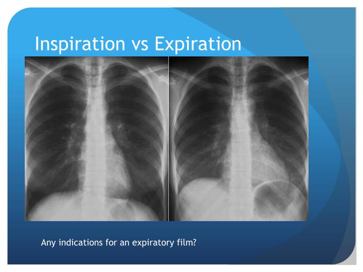
Overexposed images will have a distinct lack of quantum mottle while appearing ‘saturated’ or in extreme cases ‘burnt out’ whereby anatomy is completely obliterated from the radiograph. Underexposure errors often occur at the radiographers ends, choosing an inappropriately low exposure (low mAs) for a patient’s examination, or an examination type on the workstation.ĭue to the high dynamic range in digital imaging, overexposure is slightly more challenging to identify. In the clinical context, an underexposed chest x-ray will appear 'grainy,' and display poor penetration of the mediastinal structures leading to an inaccurate representation of anatomy. Underexposed images are easy to identify, they contain quantum mottle (noise), appear under-penetrated and often are deemed to be undiagnostic. In contemporary practice, digital radiography has replaced film technology, and with that, a more forgiving, higher dynamic range 3.


#DIRECT EXPOSURE X RAY FILM SERIES#
Put simply dynamic range is the series of exposure values that will result in a radiographic image narrow dynamic range equals a smaller window of optimal exposures 2. With practice, good quality equipment, and a few tips for adjusting your settings… Getting a properly exposed image doesn’t have to be hard!Īnd getting the right exposure will help you save time, avoid the headaches of struggling with poor quality images, and get to your diagnoses faster.ĭisclaimer: This article is for general informational purposes only, and not intended as a guide to the medical treatment of any specific animal.Traditionally, general radiography utilized film technology with a limited dynamic range, in which under or overexposed films either develop ‘too dark’ or ‘too light' 1. Use appropriate patient restraint to minimize artifacts from movement. Keep your equipment in good repair, including routine maintenance as necessary.Ĭonsider digital radiography, which can take film development out of the equation, and shorten the waiting time between views (allowing you to adjust settings prior to taking more shots). A good technique chart will remove much of the guesswork. Use technique charts for a good starting point for exposure settings. If that happens, your settings would probably be too high based on the measurement obtained from a standing patient.Īlso, be sure to follow these general tips for producing good x-ray images… For example, when taking a lateral thorax view, the thorax may be thinner when the patient is laying on their side than it was when they were standing up. Measure patients in the position they will be in during the shot. That way, you reduce the area of the primary beam and reduce scatter radiation, giving your image an overall better quality. Typically, lighter images can be obtained by lowering the kVp or mAs.Ĭheck for errors in technique, such as using or not using a grid setting.Ĭollimate. Here are some tips for getting a better shot the next time…Īdjust your settings. However, if that doesn’t work, you will probably need to retake the radiograph. Using a “hot light.” Sometimes, an overexposed radiograph can still be read by holding it up to an extra bright light known as a “hot light.” To decrease your radiograph procedure time and minimize x-ray exposure to patients and staff, it’s best to get your shots right the first time. Too short of a distance between the x-ray source and the film or plate.Īlso, scatter radiation can contribute to darkening an already over-exposed image. Reasons why your radiographs might be over-exposedĪn error in technique (kVp or mAs settings). In other words, over-exposure could cause you to miss important lesions or interfere with your interpretation of normal anatomical structures. Some structures, such as small blood vessels in the thorax, might not be visible at all.

So, rather than seeing information about the patient, you end up with a blackened image with not nearly enough detail. Over-exposed radiographs can make your life difficult because of the obliteration of normal anatomyĪn over-exposure usually means the x-ray beam was too powerful, and that a larger percentage of x-rays passed through the patient’s tissues to reach the film or plate. Instead, the image may appear “burnt out,” and overly dark or black, making it difficult to see all the information you need to make your diagnosis. This is basically the opposite of an overly white or light radiograph.
#DIRECT EXPOSURE X RAY FILM HOW TO#
In the last blog post, we talked about under-exposed radiographs and how to get better results.īut, it’s important to remember that overcompensating too far in the other direction can give you a problem in the other extreme: that is, over-exposed images.


 0 kommentar(er)
